Exploring the Complex Tissues of the Liver
Intro
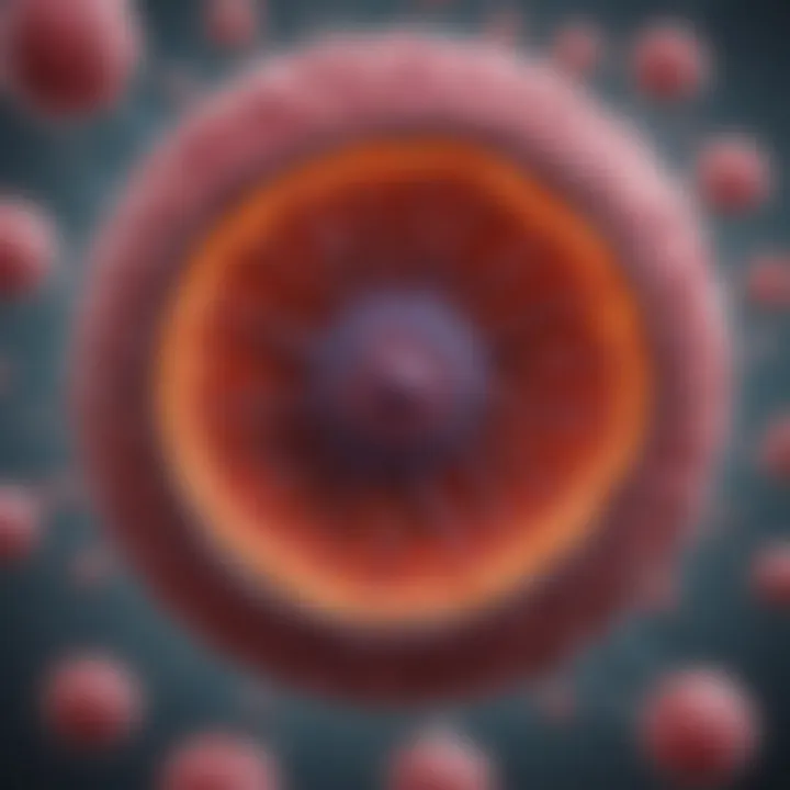
The liver is an organ that wears many hats, contributing significantly to the body's metabolic, detoxification, and regulatory activities. It’s not just a place where some things happen; it’s a bustling hub of biochemical processes, intricately woven with different types of tissues. Each tissue contributes to the liver's ability to function effectively and regenerate when needed. Understanding the complexities of these tissues—like hepatocytes, Kupffer cells, and the sinusoidal endothelium—gives us valuable insight into hepatic health and the implications of liver diseases.
In this article, we will investigate the structure and function of the various tissues within the liver. We will explore how these tissues communicate and work together, ensuring the liver maintains its role in metabolic harmony and detoxification. Also, we’ll discuss the liver’s fascinating capability to regenerate and what this means for overall health and when things go awry.
Methodology
Study Design
The primary approach to examining liver tissues involved an extensive review of existing histological studies and metabolic functions documented in scientific literature. This includes insights from both clinical research and laboratory-based studies focusing on the cellular composition of the liver. Data was distilled from peer-reviewed articles, medical journals, and histology textbooks to gather a comprehensive understanding of liver tissue.
Data Collection Techniques
Data on liver tissues were collected through:
- Histopathological Analysis: Microscopic examination of liver biopsies allows for detailed observation of the architecture and cellular components.
- Biochemical Assessments: Analysis of liver enzymes and metabolites provides insight into the functional state of hepatocytes.
- Immunohistochemistry: This technique identifies specific proteins in cells that can highlight the prevalence and state of various cell types, such as Kupffer cells and endothelial cells.
- Literature Review: In-depth exploration of studies that specifically address liver regeneration, disease impacts, and tissue interactions contributes to the overall understanding of how these tissues operate under normal and pathological conditions.
"The liver is a remarkable organ with an unparalleled ability to regenerate, often restoring its functional capacity after injury or disease."
Discussion
Interpretation of Results
The examination of liver tissues has revealed a coordinated interaction between different cell types. Hepatocytes, the primary functional cells, perform metabolic tasks and also play a crucial role in hepatocyte proliferation during liver regeneration. Kupffer cells, acting as fixed macrophages, are vital in immune responses and help clear pathogens, ensuring that the liver’s environment remains conducive to metabolic activity. Similarly, the sinusoidal endothelium acts as a filter and barrier, aiding in the regulation of the substances that enter and exit the liver.
Limitations of the Study
While this article provides substantial insights, certain limitations should be considered. For instance, the variation in liver tissue response due to factors like age, sex, and underlying health conditions could influence both the structure and function of liver tissue. Additionally, many studies focus on specific aspects of liver tissue, which may limit the broader applicability of findings. This means more holistic, multidisciplinary studies are needed for a comprehensive understanding of the liver in health and disease.
Future Research Directions
Future research should aim to:
- Investigate the genetic and molecular basis of liver tissue composition and functionality.
- Explore the roles of emerging cell types in the liver, such as newly identified stromal cells, and how they contribute to liver disease.
- Enhance understanding of liver regeneration through regenerative medicine and its potential therapeutic applications in liver diseases.
- Evaluate the effects of lifestyle factors on liver health and regeneration capabilities.
As we delve deeper into the intricate world of liver tissues, we unlock the doors to better diagnostic and therapeutic strategies that could improve outcomes for a range of liver-related conditions and enhance our understanding of human physiology.
Intro to Liver Anatomy
Understanding the anatomy of the liver serves as a foundational step for anyone studying or working in the field of liver health. The liver, the largest internal organ, plays a myriad of roles in the body, ranging from metabolism to detoxification. It is crucial to comprehend the layout and function of its tissues to grasp how various conditions can influence overall health. From the efficient processing of nutrients to the all-important filtration of toxins, the liver's architecture reveals a complex interplay that underlines its significance.
General Overview of the Liver
The liver, a reddish-brown organ, typically weighs about three pounds. It is situated under the right rib cage and is divided into two main lobes, each further segmented into lobules. Each lobule acts as a functional unit, facilitating blood flow and maintaining the liver's numerous essential functions. It's the body's master chemist, tirelessly working behind the scenes to ensure balance and health.
The organ contains a unique arrangement of cells and tissues working cohesively. At the core are hepatocytes–the predominant cells responsible for metabolic activities. Surrounding them are sinusoidal endothelial cells and specialized immune cells like Kupffer cells, each adding intricate layers of function and regulation.
"The liver doesn’t just filter; it orchestrates biochemical symphonies essential for survival."
In addition to these, the liver has a supportive extracellular matrix that aids in maintaining structure and signaling. This complex organization ensures that the liver can effectively perform its numerous duties. Understanding these elements forms the backbone of knowledge regarding liver health and disease.
Functional Importance of Liver Tissues
Each type of tissue in the liver contributes substantially to its ability to function properly. For instance:
- Hepatocytes: These cells are the workhorses of the liver, efficiently managing processes such as gluconeogenesis, lipid metabolism, and detoxification. They regulate blood sugar levels, produce bile, and manage the storage of vitamins and minerals, highlighting their metabolic prowess.
- Sinusoidal Endothelium: The unique structure of these cells allows for the filtration of blood surrounding the hepatocytes. This filtration is crucial for nutrient absorption and detoxification, yet it also ensures that immune surveillance can occur effectively.
- Kupffer Cells: Being the liver's resident macrophages, they play a key role in the immune response. They clear pathogens and debris from the blood, which helps maintain a healthy internal environment and prevent infections from taking root.
- Stellate Cells: Situated within the liver, these cells are pivotal for vitamin A storage and play a role in liver fibrosis and regeneration when the organ is injured.
In summary, understanding the anatomical makeup and functional roles of liver tissues is vital for recognizing how dysfunctions can lead to diseases like fatty liver disease, cirrhosis, or hepatitis. The liver's multifaceted operations underscore the importance of each tissue type in maintaining health and combating disease.
Types of Tissues in the Liver
Understanding the various tissue types in the liver is essential for appreciating how this organ functions and maintains homeostasis within the body. Each tissue type possesses distinct cellular components and roles, contributing to myriad physiological processes. From metabolism to detoxification, these tissues interrelate in a finely-tuned orchestration, ensuring the liver's effectiveness in its myriad functions. Knowing about these tissues aids in comprehending liver pathology and regeneration, thus underscoring its significance in both health and disease.
Hepatocytes: The Functional Units
Structure of Hepatocytes
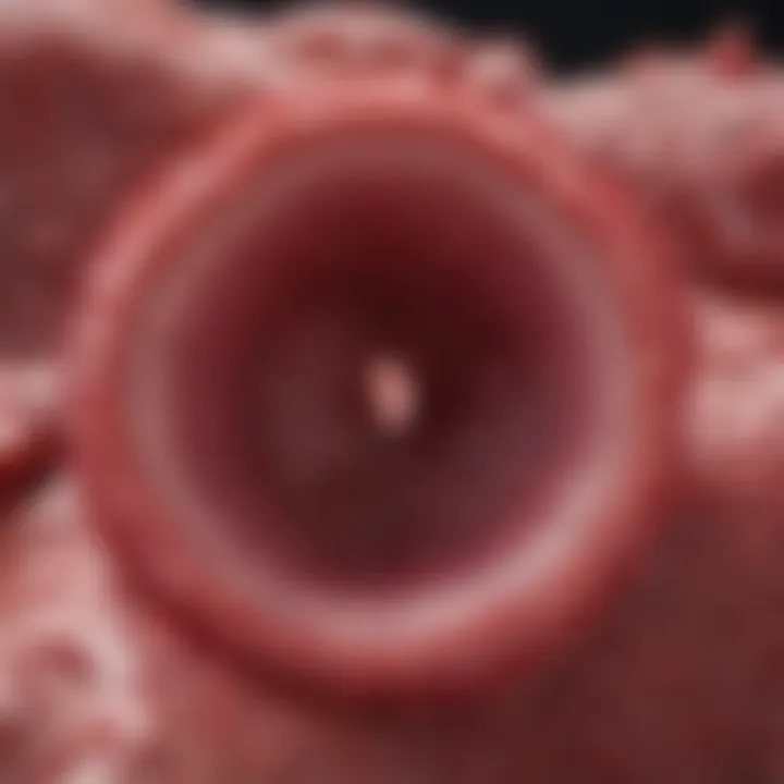
Hepatocytes are the primary functional units of the liver, making up about 70-80% of its total mass. Structurally, they are polygonal-shaped cells that exhibit a unique arrangement, enabling them to maximize contact with blood in the space of Disse. One of their most important features is the presence of numerous lipid droplets and glycogen granules, which serve as reservoirs for energy and metabolic substrates. Their extensive microvilli enhance the surface area, facilitating efficient nutrient absorption and secretion. This structural configuration is beneficial as it supports the hepatocytes' multiple functions, including protein synthesis and bile production. However, their complex architecture can also make them vulnerable to injury under pathological conditions.
Metabolic Functions of Hepatocytes
Hepatocytes are powerhouses of metabolism, responsible for a wide range of biochemical reactions including gluconeogenesis, protein synthesis, and the detoxification of drugs and toxins. One key characteristic of these cells is their ability to convert ammonia, a toxic byproduct of protein metabolism, into urea, which is then excreted by the kidneys. This metabolic ability makes them indispensable for maintaining the nitrogen balance in the body. Additionally, hepatocytes can undergo fatty acid oxidation, a crucial process for energy production. However, in conditions such as fatty liver disease, these functions can be compromised, leading to severe consequences for overall health.
Sinusoidal Endothelium: The Interface
Characteristics of Sinusoidal Endothelial Cells
The sinusoidal endothelium constitutes the blood capillaries of the liver, characterized by their fenestrated structure. This unique characteristic allows for the efficient exchange of substances, such as nutrients and metabolites, between the blood and the liver tissue. These endothelial cells are more permeable than typical capillary endothelium, enabling larger molecules to pass through. Their disposition supports liver function by maintaining a delicate balance between facilitating nutrient uptake while preventing extensive leakage of proteins. However, if their barrier integrity is compromised, it can result in significant issues, including the development of liver fibrosis.
Role in Filtration
Sinusoidal endothelial cells play a critical role in the filtration process. They help to trap pathogens and large particles from the blood, which are then cleared by the macrophages, specifically the Kupffer cells. This filtration is essential for maintaining hepatic health as it prevents the systemic spread of potential threats. Furthermore, the sinusoidal structure enhances blood flow through the liver, allowing for better filtration dynamics. A downside, however, could be the susceptibility of this intricate system to damage, leading to hepatic insufficiency or immune dysregulation over time.
Kupffer Cells: The Immune Sentinels
Nature and Function of Kupffer Cells
Kupffer cells are specialized macrophages located in the liver, acting as the first line of defense against systemic infections. They are pivotal in phagocytosing pathogens and dead cells, thus maintaining hepatic integrity. Their unique positioning within the sinusoids enables them to closely monitor the blood supply, reacting quickly to potential threats. This characteristic makes them crucial in the hepatic immune response, allowing rapid adaptation to changing conditions. One major advantage of Kupffer cells is their ability to secrete cytokines, which modulate the immune response. However, excessive activation can lead to chronic inflammation, contributing to liver diseases such as cirrhosis.
Impact on Hepatic Immunity
Kupffer cells significantly influence hepatic immunity through their interaction with lymphocytes and other immune cells. By presenting antigens and producing cytokines, they orchestrate a multifaceted immune response. Their ability to differentiate between pathogens and harmless antigens enhances immunological tolerance in the liver. This is particularly crucial given the liver's role in processing dietary substances that may otherwise trigger immune reactions. Nonetheless, an overactive response from these cells can lead to steatohepatitis, highlighting the fine balance required in immune regulation within the liver.
Stellate Cells: Storage and Regulation
Function in Vitamin A Storage
Hepatic stellate cells, also known as Ito cells, play a crucial role in the storage of vitamin A and the modulation of liver fibrosis. They are primarily located in the space of Disse. A distinct feature of these cells is their ability to store vitamin A in retinyl ester form, making them a vital part of metabolic processes related to vision and cellular health. Their storage function is beneficial as it provides a reserve that can be mobilized when needed for various cellular functions. However, excessive accumulation of lipids within these cells can lead to their activation in fibrosis.
Role in Fibrosis and Regeneration
Stellate cells become activated in response to liver injury, transforming into myofibroblast-like cells that produce extracellular matrix components. This activation is essential for wound healing but can lead to excessive scarring and fibrosis if unchecked. They also secrete growth factors that help in cellular regeneration. The delicate balance between healing and fibrosis illustrates their dual role; they are essential for recovery post-injury but can also contribute to chronic conditions if their regulation is altered.
Extracellular Matrix: Supportive Framework
Composition of the Extracellular Matrix
The extracellular matrix (ECM) in the liver is composed of a complex network of proteins and carbohydrates that provides structural and biochemical support to the liver cells. Key components include collagen, fibronectin, and glycosaminoglycans, which collectively maintain the architecture of the liver while also playing roles in cell signaling and regeneration. This matrix forms the scaffold that not only supports cellular organization but also acts as a reservoir of growth factors and cytokines essential for cellular functions. However, changes in ECM composition can lead to fibrosis, impacting liver function.
Influence on Hepatocyte Behavior
The extracellular matrix significantly influences hepatocyte behavior, including proliferation, differentiation, and response to injury. Various signals from the ECM guide hepatocyte activities, ensuring they adapt adequately to the body’s demands. An important feature of this interaction is its role in modulating liver regeneration after injury, where ECM components can either promote or inhibit regeneration depending on their composition. A disadvantage, though, could arise from an altered ECM environment leading to incorrect signaling, which may result in malignant transformations or chronic liver diseases.
Hepatic Architecture
Hepatic architecture encapsulates the structural organization and spacing within the liver, which is crucial for its diverse functions. Understanding this architecture is essential for appreciating how the liver operates, from metabolic processing to detoxification. The liver, being the body's largest internal organ, exhibits a complex arrangement that promotes efficiency in blood flow and cellular interactions. It highlights how regional specialization within the liver optimizes the varied functions required for overall health and homeostasis.
Lobular Structure of the Liver
Description of the Liver Lobule
The liver lobule is the primary functional unit of the liver, serving as a miniature processing plant for blood and nutrients. Each lobule is roughly hexagonal in shape and consists of plates of hepatocytes arranged around a central vein. This structure facilitates efficient processing of blood from the hepatic portal vein and the hepatic artery, which brings oxygen-rich blood to nourish liver cells. One of the key characteristics of the liver lobule is its structured organization, which allows for maximized interaction between hepatocytes and blood flow.
What sets the liver lobule apart is its uniqueness. Unlike many other organs, the liver’s lobules function as independent entities—each can function autonomously while still contributing to the larger organ. This is beneficial in that it allows for localized responses to various stimuli, such as hormonal regulation or toxic invasions. However, it also poses challenges, as lobules may compensate differently in cases of damage or disease, leading to uneven functional impairments across the liver.
Blood Flow within the Lobule
Blood flow within the liver lobule is a vital aspect of its architecture that plays a significant role in nutrient absorption and metabolic processes. Blood enters through the portal triad consisting of the portal vein, hepatic artery, and bile duct. The system is designed so that blood flows actively between the hepatocyte sheets as it moves toward the central vein. A key characteristic of this flow is its dual supply; the design allows the liver cells access to both nutrient-rich and oxygen-saturated blood.
The unique feature of this blood flow pattern is that it encourages thorough mixing of nutrients, hormones, and waste products, promoting an efficient metabolic environment. The advantages here are significant: it ensures that every hepatocyte has what it needs for crucial processes, like detoxification and energy production. Conversely, disruptions to this flow can create bottlenecks leading to ischemic conditions, influencing the overall health of the liver.
Portal Triads: Blood Supply and Bile Ducts
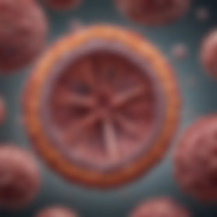
The portal triad is integral to the proper function of the liver. It is comprised of the portal vein, hepatic artery, and bile duct, forming a trinity that sustains liver activity. The connection between these three components is paramount; the portal vein carries nutrient-rich blood from the gastrointestinal tract, while the hepatic artery delivers oxygenated blood. The bile duct then transports bile produced by the liver to assist in digestion.
This sophisticated arrangement allows the liver to efficiently manage blood flow and nutrient processing, fostering an environment where metabolic regulation is finely tuned. Without the function of the portal triads, the liver would struggle to maintain its multifaceted roles within the body.
Apart from just functionality, the portal triad also presents a clear illustration of how interconnections support the overall dynamics of liver health. The architecture here not only influences how blood and bile circulate but also highlights the pathways of communication between liver cells that control diverse biochemical reactions.
The overall structural dynamics of hepatic architecture underline the complexity and elegance of liver tissues, illustrating how every element—be it cells, blood vessels, or ducts—works in harmony to maintain health and support the myriad functions of this vital organ.
Liver Regeneration Mechanisms
Liver regeneration mechanisms stand as a remarkable feature of the human body, allowing the liver to recover from injury and maintain its crucial functional balance. Understanding how this regeneration occurs is vital as it underpins the liver's role in overall metabolic health. The liver, being the body's detoxification hub, has a unique capacity to regenerate tissue, which can be seen as a blessing and a critical point of focus when analyzing liver health and pathologies.
Understanding Liver Regeneration
Phases of Liver Regeneration
Liver regeneration unfolds in a series of well-defined phases. Initially, there's the loss phase, where damage or partial resection triggers the body's innate mechanisms to initiate recovery. This is followed by the compensatory phase, where surviving hepatocytes enter a state of cell proliferation to compensate for the lost tissue. During this phase, the liver may restore its mass without fully recovering the original architecture, which can lead to complications if not monitored.
The key characteristic of the phases of liver regeneration lies in its balance between cell growth and cell death. This balance makes it a beneficial area of study, particularly for professionals focused on liver diseases and transplantations. Each phase has its unique signaling pathways and cellular responses that offer insights into how the liver can heal itself and the potential pitfalls that can arise from an imbalance.
The unique feature of these phases is that they can vary significantly based on the underlying cause of liver damage—be it viral hepatitis, acute injury, or chronic disorders. This variability presents both advantages, in terms of tailored treatment approaches, and disadvantages, as it complicates the clinical understanding of liver recovery dynamics.
Cellular Contributions to Regeneration
Cellular contributions to liver regeneration are primarily driven by hepatocytes, but several other cell types also play vital roles in this intricate process. Hepatocytes are the main protagonists, responsible for the liver’s metabolic functions, but they don't act alone. Other supportive cells like Kuppfer cells and stellate cells contribute significantly to the regeneration landscape.
A key characteristic of cellular contributions to regeneration is the interplay among different cell types. This holistic approach ensures a comprehensive recovery process, making it a popular topic for exploration in medical research and practice. Understanding how these diverse cells communicate and interact sheds light on revolutionary treatments that could aid regeneration.
One unique feature of these cellular dynamics is their plasticity—the ability to adapt and respond to varying degrees of injury significantly impacts recovery times and success rates. For instance, failing to adequately engage supporting cells in regenerative efforts can lead to dysfunctional healing, making it critical to consider these interactions in therapeutic strategies.
Factors Influencing Regeneration
Liver regeneration is not merely a mechanical response to tissue loss; it is modulated by various factors that can enhance or hinder the process effectively.
Growth Factors
Growth factors are crucial signaling molecules that directly influence the rates and efficiency of regeneration. When liver tissue is damaged, growth factors like HGF (Hepatocyte Growth Factor) are released, prompting hepatocytes to enter the cell cycle and proliferate. The role of growth factors in this process cannot be overstated, as they offer potential avenues for amplification of regenerative responses through therapeutic interventions.
The pivotal characteristic of growth factors lies in their capacity to orchestrate cell signaling pathways that regulate proliferation and survival. This makes it a beneficial choice for professionals seeking to explore regenerative medicine tactics that could significantly enhance patient outcomes. For example, recombinant growth factors are being studied for therapeutic applications following liver surgeries or injuries.
Yet, while growth factors present unique advantages in terms of enhancing recovery, there can be significant disadvantages. If growth factors are not properly controlled, excessive proliferation can lead to complications, including tumorigenesis.
Cytokines in Regenerative Processes
Cytokines play an equally important role in the regenerative processes of the liver. These small signaling proteins are produced by various cells in the liver, including Kupffer cells. They regulate immune responses and are integral in managing inflammation during the regeneration process.
A key characteristic of cytokines in regenerative processes is their capacity to create both pro-inflammatory and anti-inflammatory responses that dictate how the liver heals. This duality is beneficial, particularly in managing hepatic conditions, but it can also complicate the overall recovery process if inflammation is not properly modulated.
The distinctive feature of cytokines is that they provide a clear link between immunity and regeneration. For instance, dysregulation in cytokine signaling can lead to chronic inflammation, which can subsequently impair liver regeneration and result in fibrosis or cirrhosis. Understanding the balance and interplay of various cytokines offers fertile ground for future research aimed at harnessing these signals for therapeutic applications.
"The liver’s ability to regenerate is both a marvel of biological precision and a reminder of the importance of pathways that govern cell growth and healing."
In summary, examining liver regeneration mechanisms highlights the intricate dance of cellular responses, signaling pathways, and the myriad of factors that influence recovery. This multi-faceted understanding not only enriches our knowledge but also opens doors to innovative treatment methodologies aimed at preserving and enhancing liver health.
Pathological Changes in Liver Tissues
Understanding pathological changes in liver tissues is pivotal in the realm of hepatology, as it reveals how various diseases can alter both structure and function of the liver. This section dissect the impact of liver diseases, shedding light on how they disrupt the delicate balance of hepatic operations. Studying these pathological changes provides insights that are essential not only for the diagnosis and treatment of liver diseases but also for understanding the underlying mechanisms of liver function.
Diseases Affecting Hepatocytes
Liver Cirrhosis
Liver cirrhosis is primarily characterized by fibrosis, which is the excessive accumulation of scar tissue in the liver. This condition often stems from chronic damage due to alcoholism, hepatitis, or non-alcoholic fatty liver disease. Cirrhosis is a chief topic of consideration in this article because it epitomizes the devastating consequences of sustained liver injury. Its recognition is vital; developing cirrhosis can lead to liver failure and other life-threatening complications.
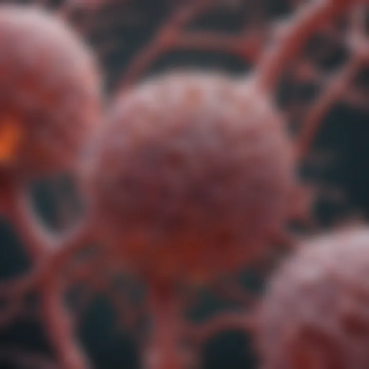
A distinctive feature of cirrhosis is the transformation of the liver from a flexible and functional organ into a hardened mass, losing its ability to perform essential detoxification and metabolic tasks efficiently.
Key Characteristics of Liver Cirrhosis:
- Formation of nodules and scar tissue
- Impaired blood flow within the liver
- Increased risk of complications such as portal hypertension and liver cancer
This disease highlights the importance of early intervention and continuous monitoring to mitigate the irreversible damage it can cause. Although advanced cirrhosis is difficult to manage, understanding its progression provides valuable information for research into potential treatments and prevention strategies.
Hepatitis
Hepatitis, whether caused by viral infections, autoimmune response, or toxins, results in inflammation of liver tissues. This inflammation can lead to significant liver dysfunction and, if left untreated, can progress to cirrhosis or liver cancer. Hepatitis is a significant focus in this article due to its prevalence and the variations it presents across different populations—chronic viral hepatitis is a global health concern.
A hallmark of hepatitis is its ability to instigate an immune response that may cause substantial tissue damage. For example, in the case of Hepatitis B and C, chronic infection can lead to both acute inflammation and long-standing tissue changes.
Unique Features of Hepatitis:
- Causes diffuse inflammation throughout the liver
- Can be either acute or chronic, with varied clinical implications
- High potential for sequelae, including cirrhosis and hepatocellular carcinoma
The identification and understanding of hepatitis are integral in devising therapeutic pathways. Importantly, vaccination against Hepatitis A and B has significantly reduced incidences, yet ongoing awareness and education are essential in managing public health concerning this illness.
Inflammatory Responses in Liver Tissues
Role of Kupffer Cells in Inflammation
Kupffer cells, the resident macrophages of the liver, play an essential role in orchestrating the immune response within hepatic tissues. They are the first line of defense against pathogens entering through the portal circulation. In this article, the discussion surrounding Kupffer cells is critical as they help maintain homeostasis while also contributing to liver pathology during disease states, such as hepatitis or cirrhosis.
Key Characteristics of Kupffer Cells:
- Phagocytose pathogens and apoptotic cells
- Secrete a range of inflammatory cytokines
- Contribute to the regulation of immune responses
The unique ability of Kupffer cells to both mediate inflammation and facilitate liver repair during acute insults stands as a double-edged sword. Excessive activation may lead to chronic inflammation, further exacerbating liver diseases.
Impact of Chronic Inflammation on Liver Health
Chronic inflammation in the liver significantly disrupts its normal architecture and function. From steatosis to cirrhosis, an array of chronic conditions arise from prolonged inflammatory reactions. Understanding this dynamic is vital for addressing the broader implications that chronic inflammation has on the liver.
Key Characteristics of Chronic Inflammation:
- Continuous injury to liver cells
- Can lead to cellular apoptosis and necrosis
- Often associated with fibrotic changes over time
This connection between chronic inflammation and liver diseases further reinforces the need for ongoing research in therapeutic interventions aimed at moderating the inflammatory processes of the liver.
Fibrosis and the Role of Stellate Cells
Stellate cells are crucial players in the development of liver fibrosis. Upon liver injury, they become activated and transition from a quiescent state to a fibrogenic state, producing collagen and other extracellular matrix components that contribute to scar tissue formation. This topic is of great importance as it provides key insights into the mechanisms behind fibrosis—a condition that can lead to cirrhosis and liver failure over time.
How Stellate Cells Contribute to Fibrosis:
- Produce collagen in response to liver injury
- Their activation is tightly regulated by various cytokines
- Play a role in regenerating tissue after injury
The balance between fibrosis and regeneration orchestrated by stellate cells is what defines hepatic health, making the study of their function not just interesting but also essential for devising new treatments for liver diseases.
Ending: The Complexity of Liver Tissues
In exploring the rich landscape of liver tissues, we find a tapestry woven from a variety of specialized cells that each hold distinct roles crucial to the liver’s multifaceted functions. This section underscores the importance of understanding these tissues—not just as isolated components, but as part of a grander system that harmonizes metabolism, detoxification, and immune response. The complexity of liver tissues fosters immense adaptability and resilience, making it a fascinating organ to study.
Integrating Structure and Function
The architecture of liver tissues is intricately designed to support its diverse functions. Each tissue type—be it hepatocytes, Kupffer cells, or sinusoidal endothelium—wasn't merely thrown together but instead reflects a thoughtfully organized synergy where structure complements function.
- Hepatocytes, the workhorses of the liver, are uniquely structured with a large surface area to optimize metabolic activity.
- Kupffer cells, acting as sentinels of the immune system, reside strategically within the sinusoids, skillfully positioned to capture pathogens circulating in the blood.
- Sinusoidal endothelial cells, with their porous nature, work in concert to facilitate nutrient absorption and waste removal, showcasing a blend of filtration capabilities and vascularization.
The relationship between structure and function in the liver exemplifies biological optimization. Understanding how these elements integrate can inform research in regenerative medicine and liver disease treatment.
Implications for Future Research
The exploration of liver tissues brings forth numerous avenues for future inquiry. As our knowledge deepens regarding the complexities of liver structure and function, several key areas emerge as ripe for investigation:
- Pathway Disruptions: Research into how specific disruptions in liver tissues contribute to diseases like cirrhosis and hepatocellular carcinoma can lead to novel therapeutic strategies.
- Regenerative Mechanisms: Understanding the molecular signals that govern liver regeneration can unlock potential treatments for acute liver damage.
- Immune Dynamics: Exploring the role of Kupffer cells and other immune components in chronic liver diseases presents opportunities to enhance immunotherapeutic interventions.
"The liver is often termed the body’s chemical processing plant, and dissecting its complexities provides insights not only into liver health but overall well-being."
In summary, as we close this examination of liver tissues, it becomes evident that their intricacies reflect both evolutive adaptations and functional imperatives. Continuous research is essential to further unravel these complexities, heralding new understanding and improved outcomes in hepatic health and disease management.


