Cerebral Pontine Angle Tumors: An In-Depth Review


Intro
Cerebral pontine angle tumors are a specialized group of tumors located at the intersection of the brainstem and cerebellum. The complexity of their anatomy and the variety of tumor types complicates both diagnosis and treatment. Understanding these tumors is essential for neurologists, neurosurgeons, and oncologists alike. This article aims to provide a deep dive into the underlying biology, clinical manifestations, diagnostic techniques, treatment modalities, and prognostic outcomes associated with these tumors.
Each section of this article will highlight key points and contemporary advancements in research, thereby offering a comprehensive resource for students, educators, and medical professionals to better understand cerebral pontine angle tumors.
Methodology
Study Design
This review endeavors to synthesize current literature on cerebral pontine angle tumors. By assessing peer-reviewed studies, clinical trials, and case reports, we aim to provide a multifaceted perspective on this domain of neuro-oncology. The design integrates both quantitative and qualitative data to ensure a well-rounded understanding.
Data Collection Techniques
Data collection for this article included:
- Literature Review: A thorough examination of recent studies related to diagnosis, treatment options, and outcomes for cerebral pontine angle tumors.
- Clinical Trial Databases: Data from ongoing and completed trials were evaluated to discuss emerging therapies and techniques.
- Expert Interviews: Insights from leading professionals in the field of neurosurgery and oncology were also factored into the analysis.
Discussion
Interpretation of Results
The results suggest a pressing need for a more standardized approach in both diagnostics and treatment. Various tumor types, such as schwannomas and meningiomas, present unique challenges. Early diagnosis often leads to better patient outcomes. Multimodal approaches, combining surgery and radiotherapy, are shown to be effective, though controversial in some cases.
Limitations of the Study
While this article strives for comprehensiveness, there are limitations. The heterogeneity of tumor types results in variability in treatment responses. Furthermore, the sources reviewed may be subject to bias, as some studies have smaller sample sizes or lack long-term follow-up.
Future Research Directions
Future research in this field could focus on:
- Molecular Biology: Identifying genetic markers specific to cerebral pontine angle tumors may lead to tailored therapeutic approaches.
- Improved Diagnostic Tools: Developing advanced imaging techniques could facilitate earlier and more accurate diagnoses.
- Longitudinal Studies: Long-term follow-up studies could provide better insight into prognosis and survivorship.
By systematically examining cerebral pontine angle tumors, we hope to contribute to improved clinical practice and better outcomes for patients.
Prologue to Cerebral Pontine Angle Tumors
Cerebral pontine angle tumors occupy a significant position within the study of neuro-oncology. Understanding these tumors provides critical insights into not only their biological behavior but also their clinical implications. Their location at the junction of the brainstem and cerebellum makes them particularly intriguing as they can impact a variety of neurological functions. Hence, recognizing their types, symptoms, and treatment options is essential for both medical professionals and patients seeking clarity on this condition.
Definition and Location
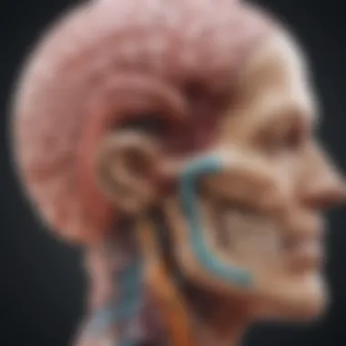
Cerebral pontine angle tumors are neoplasms that develop in the cerebellopontine angle region of the brain. This area is situated between the cerebellum and the pons, a part of the brainstem. The tumors can affect key neural structures in this region, leading to a broad spectrum of neurological symptoms. The anatomical complexity of the angle often contributes to diagnostic challenges, making precise identification crucial for effective treatment. Common types of tumors found in this location include vestibular schwannomas and meningiomas.
Historical Context
The history of cerebral pontine angle tumors is marked by evolving understanding and advancements in medical technology. Initially, these tumors were poorly understood, and surgical intervention was fraught with significant risks. With the invention of high-resolution imaging techniques like MRI in the 1980s, the ability to accurately visualize these tumors has improved significantly. Over the decades, research has expanded our understanding of their biological behavior, contributing to better outcomes for patients. Pioneers in neurosurgery have also played a vital role, developing surgical approaches that minimize complications while maximizing tumor resection. The cumulative efforts in research, diagnostics, and treatment options have made significant strides in improving patient management and outcomes.
Types of Cerebral Pontine Angle Tumors
Understanding the various types of cerebral pontine angle tumors is crucial for accurate diagnosis, treatment planning, and management of patient care. Each tumor type has distinct characteristics, behavior, and treatment considerations that significantly impact patient outcomes. A thorough appreciation of these types ensures that both medical professionals and patients can make well-informed decisions regarding therapeutic options.
Vestibular Schwannomas
Vestibular schwannomas, commonly known as acoustic neuromas, arise from Schwann cells that myelinate the vestibulocochlear nerve (CN VIII). Typically, these tumors are benign and slow-growing. Their location at the cerebellopontine angle means they can exert pressure on adjacent structures, leading to a variety of symptoms.
Common presentations include:
- Hearing loss, often progressive and unilateral.
- Tinnitus, perceived as ringing in the ear.
- Balance problems due to vestibular nerve involvement.
The management of vestibular schwannomas can vary. Observation is an option for small tumors that do not produce significant symptoms. Surgical excision or focused radiation therapy may be necessary for larger tumors or those causing symptoms. Timely consideration of these treatment options is essential to preserve auditory function and prevent complications.
Meningiomas
Meningiomas originate from the meninges, the protective membranes surrounding the brain and spinal cord. These tumors can be located in the cerebral pontine angle region, leading to varying neurological implications based on their size and location. While most meningiomas are benign, some can exhibit aggressive behavior.
Symptoms may include:
- Difficulty with coordination or balance.
- Hearing loss or speech issues, depending on the tumor's impact on nearby structures.
- Various focal neurological deficits due to compression effects.
The treatment for meningiomas typically involves surgical resection, particularly for those that cause symptoms or show growth over time. In some cases, radiation therapy may be a reasonable alternative, especially for inoperable tumors or those deemed high-risk for surgical complications.
Atypical Teratoid Rhabdoid Tumors
Atypical teratoid rhabdoid tumors (ATRTs) are rare and aggressive neoplasms mainly affecting children but can occur in adults. When these tumors appear in the cerebral pontine angle, they can cause rapid neurological decline. ATRTs are often characterized by specific genetic mutations, especially in the SMARCB1 gene.
Clinical features may include:


- Severe headaches.
- Rapidly progressing neurological deficits.
- Symptoms of increased intracranial pressure.
The management of ATRTs necessitates aggressive approaches, including surgical intervention followed by chemotherapy and radiation. Given their high malignancy potential and associated poor prognosis, early detection and comprehensive treatment planning are paramount.
Ependymomas
Ependymomas develop from ependymal cells lining the ventricles of the brain and the spinal cord. When these tumors occur in the cerebral pontine angle, they can disrupt normal brain function and present challenging symptoms. Ependymomas can be classified as either benign or malignant, with the latter often showing more aggressive behavior.
Key symptoms associated with ependymomas may include:
- Intractable headaches due to increased intracranial pressure.
- Dizziness and balance issues resulting from pressure on the brainstem.
- Signs of cranial nerve involvement, depending on the tumor's location.
Surgical resection remains the primary treatment option, and the extent of removal is often linked to patient outcomes. Adjunctive therapies, such as radiation, may follow surgery based on the tumor’s histological characteristics and the overall prognosis.
"Understanding tumor typology at the cerebral pontine angle is essential for effective management strategies and tailoring patient care."
Each of these tumor types has unique presentations and treatment challenges. Knowledge about them enhances understanding of patient experiences and aids healthcare providers in offering optimized care aligned with the distinct characteristics of each tumor.
Symptoms and Clinical Presentation
Understanding the symptoms associated with cerebral pontine angle tumors is crucial for timely diagnosis and effective treatment. These tumors can present a wide array of neurological manifestations that impact various functions including sensation, coordination, and hearing. Recognizing these symptoms early can lead to better management and outcomes for patients.
Common Neurological Symptoms
Cerebral pontine angle tumors often result in several common neurological symptoms. These may include:
- Headaches: Generally, these headaches can vary from mild to severe and may not respond to typical analgesics.
- Dizziness: Patients frequently report episodes of vertigo, which can severely affect their balance.
- Facial Weakness or Numbness: This can affect one side of the face, as these tumors may impact cranial nerves responsible for facial motor functions.
The presence of these symptoms can stem from increased intracranial pressure or direct encroachment on cranial nerves, thus impacting functionality. Clinicians should take a detailed medical history and conduct a thorough neurological examination to recognize these symptoms and understand their implications for cerebral health.
Cerebellar Symptoms
As these tumors are situated near the cerebellum, symptoms can significantly affect coordination and balance. Common cerebellar symptoms include:
- Ataxia: This is a condition characterized by uncoordinated movements, which can manifest as stumbling or poor balance.
- Tremors: Patients may experience involuntary shaking movements, which can affect fine motor skills.
- Difficulty with Fine Motor Tasks: Tasks that require precision, such as writing or buttoning clothes, may become significantly challenging.
Awareness of cerebellar symptoms is important as these indicate direct impacts on the brain’s ability to regulate movement and posture. Referral for imaging studies becomes necessary if such symptoms arise.
Impact on Hearing
Hearing disturbances are often a significant concern with cerebral pontine angle tumors, particularly vestibular schwannomas. These tumors can affect the vestibulocochlear nerve, leading to:
- Hearing Loss: This can be gradual or sudden, often affecting one ear more than the other.
- Tinnitus: Ringing in the ears can be a persistent issue, leading to discomfort and disturbance.
- Balance Problems: Since the vestibular system is involved in maintaining equilibrium, issues with balance might also arise.
The impact on hearing highlights the need for comprehensive audiological assessments as part of patient evaluations. Understanding these auditory changes can guide appropriate interventions that improve the quality of life for affected individuals.
In summary, recognizing the multifaceted symptoms of cerebral pontine angle tumors is essential for healthcare providers. Timely identification can facilitate early intervention, ultimately improving patient outcomes.
Diagnostic Approaches
The importance of diagnostic approaches in the evaluation of cerebral pontine angle tumors cannot be overstated. An accurate diagnosis is foundational for determining the appropriate treatment plan and assessing prognosis. Given the complex nature of these tumors, advanced methods are critical for visualizing the tumor's size, location, and impact on adjacent structures. In doing so, these approaches help in distinguishing these tumors from other possible conditions, which is a vital step in ensuring that patients receive the most effective care possible.
Imaging Techniques
Imaging techniques are among the first steps in forming a diagnosis for tumors located in the cerebral pontine angle. The two primary modalities utilized are Magnetic Resonance Imaging and Computed Tomography scans. Each technique has its strengths and weaknesses, which can influence their selection during patient assessment.
MRI
Magnetic Resonance Imaging (MRI) plays a significant role due to its superior soft tissue contrast. This imaging technique allows for a detailed view of cranial structures, making it possible to assess the tumor's characteristics effectively. A key characteristic of MRI is its ability to provide high-resolution images without the use of ionizing radiation, making it a safer choice for repeated studies in patients.
One unique feature of MRI is its capability to utilize different sequences and contrast agents, enhancing the visibility of lesions. This advantage aids in distinguishing between tumor types, which can guide therapeutic decisions. However, it is important to note that MRI may take longer than CT scans, which can be a disadvantage in certain emergency situations.
CT Scans
Computed Tomography (CT) scans are beneficial for their speed and availability, particularly in emergency settings. This imaging method is particularly useful in identifying acute bleeding or significant mass effects caused by tumors. A major highlight of CT scans is their efficiency in providing quick images of the brain and surrounding anatomy.
CT scans are unique in their capability to provide bone windows, which can be beneficial in assessing any associated bony involvement with a tumor. Nevertheless, they may not offer the same level of detail in soft tissue contrast as MRI. This limitation can sometimes hinder effective differentiation of tumor type.
Histological Examination
Histological examination is vital in confirming the diagnosis of cerebral pontine angle tumors. This process involves obtaining a tissue sample through biopsy for microscopic analysis. The examination provides information on the cellular composition of the tumor, helping classify it into specific types. Understanding the histological type contributes significantly to determining treatment strategies and predicting outcomes.
Differential Diagnosis
Differential diagnosis is a critical component of assessing cerebral pontine angle tumors. It involves distinguishing these tumors from various other conditions that can mimic their symptoms or imaging appearance. Conditions such as vestibular schwannomas, meningiomas, and other focal lesions may present similarly. A thorough differential diagnosis is essential to ensure that the patient receives appropriate management and avoids unnecessary procedures. This process often includes correlating clinical findings with radiographic features, as well as integrating laboratory results when applicable.
In summary, accurate diagnostic approaches are paramount for effectively addressing cerebral pontine angle tumors. By utilizing advanced imaging techniques, conducting histological examinations, and engaging in differential diagnosis, healthcare professionals can develop a comprehensive understanding of these tumors, ultimately leading to improved patient outcomes.


Treatment Options
The treatment options for cerebral pontine angle tumors play a crucial role in shaping patient outcomes and quality of life. Selecting the most suitable approach is vital based on tumor type, size, and patient health. This section examines various treatment modalities, emphasizing the importance of a personalized and multidisciplinary approach.
Surgical Interventions
Microsurgery
Microsurgery is a highly specialized surgical technique that allows for greater precision in operating on brain tumors. The significance of microsurgery in treating cerebral pontine angle tumors cannot be overstated. It enables surgeons to access tumors with minimal disruption to surrounding neural structures. The key characteristic of microsurgery is the use of high-powered microscopes and advanced surgical instruments that facilitate delicate dissection and tumor removal.
One of the primary benefits of microsurgery is its ability to significantly reduce recovery time and postoperative complications. By minimizing the impact on healthy tissue, patients may experience less trauma and quicker rehabilitation. Furthermore, the enhanced visualization offered by microsurgical techniques allows for a more complete tumor resection, which is essential for improving long-term prognoses.
However, there are some disadvantages associated with microsurgery. It requires highly trained surgeons with expertise in neuroanatomy, and not all tumors may be accessible through this method due to their location or adherence to critical structures.
Endoscopic Techniques
Endoscopic techniques represent an innovative approach in the field of neurosurgery, particularly for tumors located in challenging areas like the cerebral pontine angle. These techniques utilize a thin, flexible tube equipped with a camera and surgical instruments. This allows for visualization of the tumor and surrounding brain structures without the need for large incisions.
The key characteristic of endoscopic techniques is their minimally invasive nature. This makes them a popular choice for many patients. By employing these techniques, surgeons can often achieve effective tumor resection while minimizing damage to surrounding tissues. The unique feature of this approach is the ability to navigate through natural openings in the skull, such as the nose or mouth, thereby reducing recovery times and hospital stays.
Despite these advantages, endoscopic techniques also have limitations. The scope of visibility can be restricted, making it difficult to assess the entire tumor or surrounding anatomy during surgery. Additionally, the need for specialized equipment and expertise can affect accessibility for some patients.
Radiation Therapy
Radiation therapy serves as a key treatment option, especially for patients unable to undergo surgery due to various factors. This modality employs targeted radiation to destroy tumor cells or inhibit their growth. Often used as an adjunct to surgical interventions, radiation therapy can enhance treatment efficacy, particularly in cases of residual tumor following surgery. The precision of modern radiation techniques allows for the sparing of nearby healthy brain tissue, which is critical in the sensitive regions around the cerebral pontine angle.
Chemotherapy Considerations
Chemotherapy is less commonly employed for cerebral pontine angle tumors compared to other modalities. However, it may be considered for specific histological types or advanced cases. This treatment approach utilizes anticancer medications to stop tumor growth or induce cell death. One significant challenge with chemotherapy is its systemic nature, which can lead to numerous side effects. Therefore, a careful assessment must be conducted to weigh the potential benefits against the possible adverse effects. As research progresses, the development of targeted therapies may enhance the feasibility of chemotherapy in this context.
Prognostic Factors and Outcomes
Understanding the prognostic factors and outcomes associated with cerebral pontine angle tumors is critical for both patients and clinicians. Prognostic factors can influence treatment decisions and overall patient management. They help in estimating the likely outcomes based on certain key indicators. By focusing on specific elements like tumor location and size, patient age and health status, and the histological types of tumors, one can make informed decisions regarding treatment modalities and follow-ups.
Tumor Location and Size
The location and size of the tumor play a significant role in determining the prognosis for patients diagnosed with cerebral pontine angle tumors. Tumors that are positioned deeper within the cerebellopontine angle can be more challenging to access and remove surgically. Larger tumors may also lead to increased pressure on adjacent structures, exacerbating symptoms, and can complicate the surgical approach. When discussing size, tumors over five centimeters are often classified as large and typically correlate with poorer outcomes compared to smaller lesions.
- Location: Tumors located near critical neurological pathways tend to present greater surgical risks and may lead to higher complications. For example, a tumor impinging on the cranial nerves may compromise functions critical for hearing and balance.
- Size: As mentioned, larger tumors not only pose surgical challenges but also tend to lead to a greater degree of neurological impairment, affecting overall quality of life post-treatment.
A focused analysis on tumor characteristics greatly enhances the ability to predict survival rates and recovery trajectories.
Patient Age and Health Status
The age and overall health status of a patient are vital prognostic indicators when considering treatment options and outcomes in cerebral pontine angle tumors. Generally, younger patients tend to have better outcomes due to their robust health and greater physiological reserves.
- Age: Older patients may experience comorbidities that complicate treatment options. Moreover, their physiological responses to surgery or radiation therapy can differ markedly from those of younger individuals.
- Health Status: The presence of preexisting conditions, such as cardiovascular disease or diabetes, can hinder recovery. A patient's overall performance status, assessed through metrics like the Karnofsky Performance Scale, can provide insight into their ability to withstand the implications of surgery or other treatments.
Histological Type of Tumor
The specific histological type of tumor also substantially impacts prognosis. Different tumor types exhibit varied growth patterns, locational preferences, and responses to treatment, which can fundamentally alter the expected outcomes for a patient.
- Histological Variants: For instance, vestibular schwannomas often have a more favorable prognosis compared to aggressive variants like atypical teratoid rhabdoid tumors.
- Growth Rate: The growth rate of histological types influences how quickly patients may develop symptoms and seek treatment. Rapid growth can lead to increased symptoms and more urgent interventions, while slower-growing tumors might allow for a more conservative approach.
In summary, prognosis in cerebral pontine angle tumors is multifaceted, relying on several core factors such as the tumor's location and size, the patient's age and health status, and the histological type of the tumor. Focusing on these aspects enables better planning for treatment strategies and provides a clearer understanding of what outcomes can be expected. A comprehensive approach ensures personalized care, benefiting patients through tailored treatment protocols.
Challenges in Treatment
The management of cerebral pontine angle tumors encompasses considerable challenges which directly influence patient outcomes. Understanding these challenges is crucial, as they inform medical professionals about the complexities involved in treatment planning and patient care. The impact of surgical risks, complications, and the adverse effects of radiation therapy cannot be understated. Significant risks can alter the course of treatment, making it imperative to develop tailored strategies for each patient.
Surgical Risks and Complications
Surgery is often the primary treatment for cerebral pontine angle tumors. However, the unique anatomy of this region makes surgical interventions particularly risky. Surgeons must navigate through delicate brain structures, which can lead to complications such as:
- Neurological deficits: Damage to critical areas can result in long-term complications, including weakness, coordination issues, or speech problems.
- Infection: As in any surgical procedure, the risk of postoperative infection exists. Infections can complicate recovery and affect overall health.
- Hemorrhage: Uncontrolled bleeding can occur during or after surgery, posing immediate health risks to the patient.
- Cerebrospinal fluid leaks: Improper closure may result in leaks, increasing the chance of meningitis or requiring additional interventions.
These risks necessitate careful preoperative assessments and a thorough discussion with patients about the potential outcomes and limitations of surgery. Planning for postoperative care is just as crucial as the surgical procedure itself, as this determines recovery success and quality of life.
Adverse Effects of Radiation Therapy
Radiation therapy serves a significant role in the management of cerebral pontine angle tumors, either as a primary treatment or in cases where surgery is not feasible. Nonetheless, this form of treatment is not without its challenges, as it can lead to a range of adverse effects, including:
- Fatigue: Patients often experience fatigue during and after treatment, which can impact daily functioning and recovery.
- Skin reactions: The area receiving radiation may exhibit redness, irritation, or changes in texture, causing discomfort.
- Neurological side effects: Short-term effects can include headaches, nausea, and cognitive impairments. These may diminish over time but can be quite distressing during active treatment.
"Understanding the full spectrum of treatment challenges enables healthcare providers to offer more personalized and effective care."
- Long-term risks: Longitudinal studies indicate there may be an increased risk of secondary tumors or other long-term neurological deficits due to radiation exposure, depending on the cumulative dose.


These adverse effects highlight the necessity for ongoing patient monitoring and support. Careful consideration should be given to each patient’s situation, ensuring that the benefits of radiation therapy outweigh the potential risks.
Current Research and Innovations
Current research and innovations in cerebral pontine angle tumors are crucial in improving patient outcomes and advancing medical understanding in this specific area of neuro-oncology. These tumors, while relatively rare, present distinct challenges in diagnosis and treatment due to their intricate location and the critical structures nearby. Ongoing studies aim to refine surgical techniques, enhance diagnostic accuracy, and develop more effective therapeutic strategies. This not only has implications for treatment success but also fundamentally affects quality of life for patients.
Being at the forefront of this research can bring about significant advancements. The integration of new technologies and methodologies greatly influences surgical outcomes. Innovations such as robotic-assisted surgery and intraoperative imaging allow more precise tumor removal, reducing the likelihood of complications. Additionally, these approaches may improve recovery times, which is of paramount importance for the patients affected by these complex tumors.
Research into innovative therapies is equally vital. As the understanding of the molecular biology of these tumors advances, researchers are exploring targeted treatments that can enhance effectiveness while minimizing side effects. This section will detail key advancements in surgical techniques and novel therapeutic approaches, demonstrating their significance in managing cerebral pontine angle tumors.
Advancements in Surgical Techniques
Advancements in surgical techniques represent one of the most significant areas of research pertaining to cerebral pontine angle tumors. Traditional surgical methods have had limitations, often associated with complications and prolonged recovery times. Today, however, the advent of minimally invasive techniques has revolutionized the approach to tumor resection.
Surgical innovations include:
- Microsurgical techniques: These provide enhanced visualization and control, enabling surgeons to excise tumors with more precision, preserving adjacent neural structures.
- Endoscopic approaches: Utilizing narrow, flexible instruments, surgeons can access the tumor with minimal damage to surrounding tissues.
- Robotic-assisted surgeries: These surgeries enable more dexterous movements and improved ergonomics, which aid in complex cases typically associated with higher risks of complications.
Moreover, intraoperative imaging techniques such as fluorescein-guided surgery allow surgeons to visualize tumor margins better and distinguish healthy tissue from tumorous cells in real time. This reduces the chance of leaving residual tumor, thereby potentially enhancing long-term survivorship.
Novel Therapeutic Approaches
The exploration of novel therapeutic approaches to cerebral pontine angle tumors is an active area of research. Alongside surgical interventions, innovative non-surgical treatments are under investigation to improve patient outcomes.
Key areas of focus in this regard include:
- Targeted therapies: These drugs specifically attack cancer cells based on their molecular characteristics, reducing damage to normal cells and minimizing side effects.
- Immunotherapy: This treatment harnesses the body’s immune system to fight tumors. Current trials are assessing the effectiveness of immune checkpoint inhibitors and other modalities in treating these tumors.
- Gene therapy: Ongoing research examines the use of gene editing technologies to correct abnormalities at the cellular level, potentially reversing tumor growth.
New clinical trials are essential to evaluate the safety and efficacy of these innovative therapeutic approaches. The findings could lead to significant breakthroughs in managing this challenging category of tumors, which have traditionally lacked effective treatment options.
These advancements represent a beacon of hope for patients facing cerebral pontine angle tumors, as multidisciplinary strategies continue to evolve and improve.
Management of Patient Care
The management of patient care in cases of cerebral pontine angle tumors is vital for improving outcomes and enhancing quality of life. This multidisciplinary approach addresses the complexity of these tumors due to their location and potential impact on crucial neurological functions. Effective patient management integrates multiple perspectives, ensuring that patients receive comprehensive care tailored to their individual needs.
One significant element in patient care is the coordination among specialists, including neurologists, neurosurgeons, radiologists, and oncologists. This collaboration fosters a unified treatment plan that considers the tumor type, patient health, and other factors. Benefits of this approach encompass improved communication, a more accurate understanding of the patient’s condition, and the ability to adjust treatment plans as needed. As symptoms can vary significantly, a holistic view can lead to better management strategies that address not just the tumor but the overall impact on patient health as well.
Patient care also involves continuous monitoring. Regular follow-ups can help track symptoms, assess the effectiveness of treatment, and make timely adjustments if adverse effects occur. Patients often face challenges with emotional well-being following a diagnosis. Herein lies another critical aspect: integrating psychological support into the care framework. This inclusivity denotes a recognition of the multifaceted struggles patients encounter beyond just tangible medical issues.
In summary, patient care management stands as a cornerstone in addressing cerebral pontine angle tumors. By ensuring cohesive collaboration and ongoing support, healthcare providers can greatly improve patient experiences and outcomes.
Multidisciplinary Treatment Teams
Multidisciplinary treatment teams play an indispensable role in the management of patients with cerebral pontine angle tumors. These teams typically include professionals from various fields, offering a diverse array of expertise and insights. Each member contributes unique knowledge, leading to a well-rounded perspective that enhances treatment outcomes.
The existence of such teams fosters a joint decision-making atmosphere. This ensures that all potential treatment options are explored and that the chosen paths align well with the patient’s individual goals and preferences. The complexity associated with cerebral pontine angle tumors requires precision and collaboration; hence, the integration of surgeons, radiation oncologists, and rehabilitation specialists is essential.
Rehabilitation and Support
Rehabilitation and support services are critical components of effective patient management after treatment for cerebral pontine angle tumors. These services not only address physical recovery but also encompass emotional and psychological support for patients.
Speech Therapy
Speech therapy is an important aspect of rehabilitation for patients who may experience speech or communication difficulties due to their tumors or treatments. The key characteristic of speech therapy is its focus on improving communication skills, enabling patients to express themselves clearly. It is often recommended due to its tailored approach of addressing individual patient needs.
One unique feature of speech therapy is its incorporation of exercises that target the specific aspects of communication that patients struggle with. For example, this may include articulation, language comprehension, or cognitive-linguistic skills. Advantages of this therapy include improved quality of life and enhanced social interactions, while potential disadvantages may involve the time commitment required for therapy sessions.
Physical Therapy
Physical therapy also holds a significant place in rehabilitation post-treatment. Its main goal is to improve mobility and strength, minimizing any physical deficits that may arise as a result of surgery or tumor impact. The key characteristic of physical therapy is its broad approach, often employing various exercises and techniques to restore function.
A unique feature of physical therapy is its adaptability to the patient’s specific needs. It can include tailored exercise regimens, balance training, and strength-building activities. The advantages of physical therapy are notable, as many patients find significant improvements in mobility, whereas some may experience gradual progress, which requires patience and ongoing commitment.
Epilogue and Future Directions
In concluding this exploration of cerebral pontine angle tumors, it is vital to reflect on both the findings presented and the implications for future research and treatment. These tumors, located in a critical junction of the central nervous system, pose significant diagnostic and therapeutic challenges. Thus, understanding their nature is not merely academic; it directly affects clinical outcomes and patient care, influencing how practitioners approach these complex conditions.
Summary of Key Findings
This article identifies several significant insights regarding cerebral pontine angle tumors. The types discussed include vestibular schwannomas, meningiomas, atypical teratoid rhabdoid tumors, and ependymomas, each presenting unique challenges in terms of symptoms and treatment options.
Key findings include:
- Variety in Tumor Types: The diverse histological characteristics of these tumors necessitate customized treatment protocols.
- Impact on Quality of Life: Symptoms such as hearing loss and balance issues can substantially affect a patient's daily functioning and psychological well-being.
- Advances in Diagnostics and Treatment: Technological advancements in imaging and surgical techniques have improved the ability to detect and treat these tumors effectively.
- Multidisciplinary Management: Successful management often involves a coordinated approach among various medical specialists, ensuring comprehensive care.
Potential Areas for Future Research
Future research endeavors must focus on several critical areas:
- Molecular Studies: Investigating the molecular genetics of these tumors to uncover targeted therapies may lead to more effective treatments.
- Long-Term Outcomes: More studies tracking long-term outcomes of various treatment modalities can provide insights into efficacy and quality of life post-treatment.
- Innovative Surgical Techniques: Ongoing refinement of surgical methods, including robotic-assisted surgeries, could improve precision and reduce recovery times.
- Health Economics: Analyze the cost-effectiveness of different treatment strategies, which will aid in resource allocation and patient accessibility to care.
In summary, while the current understanding of cerebral pontine angle tumors has advanced significantly, there remains ample opportunity for exploration and innovation in the field. By prioritizing research in these targeted areas, medical professionals can enhance outcomes and improve the overall management of cerebral pontine angle tumors.


